Petrography
Our standard approach - We get lots of mileage out of it.

Our standard approach - We get lots of mileage out of it.
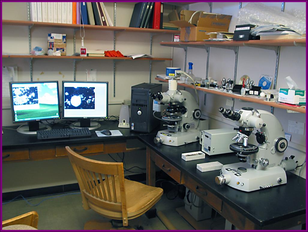
Two Zeiss Photo 3 petrographic microscopes with reflected light, epifluorescence, phase contrast, Nomarsky, and integrated 35 mm film cameras.
Interfaced with Pixera 6 megapixel digital camera and PC-based image collection and processing.
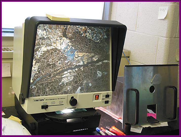
Petroscope projection macroscope. Allows viewing of entire thin section for larger context of small scale petrographic features. Also good for quick screening of thin section suites.
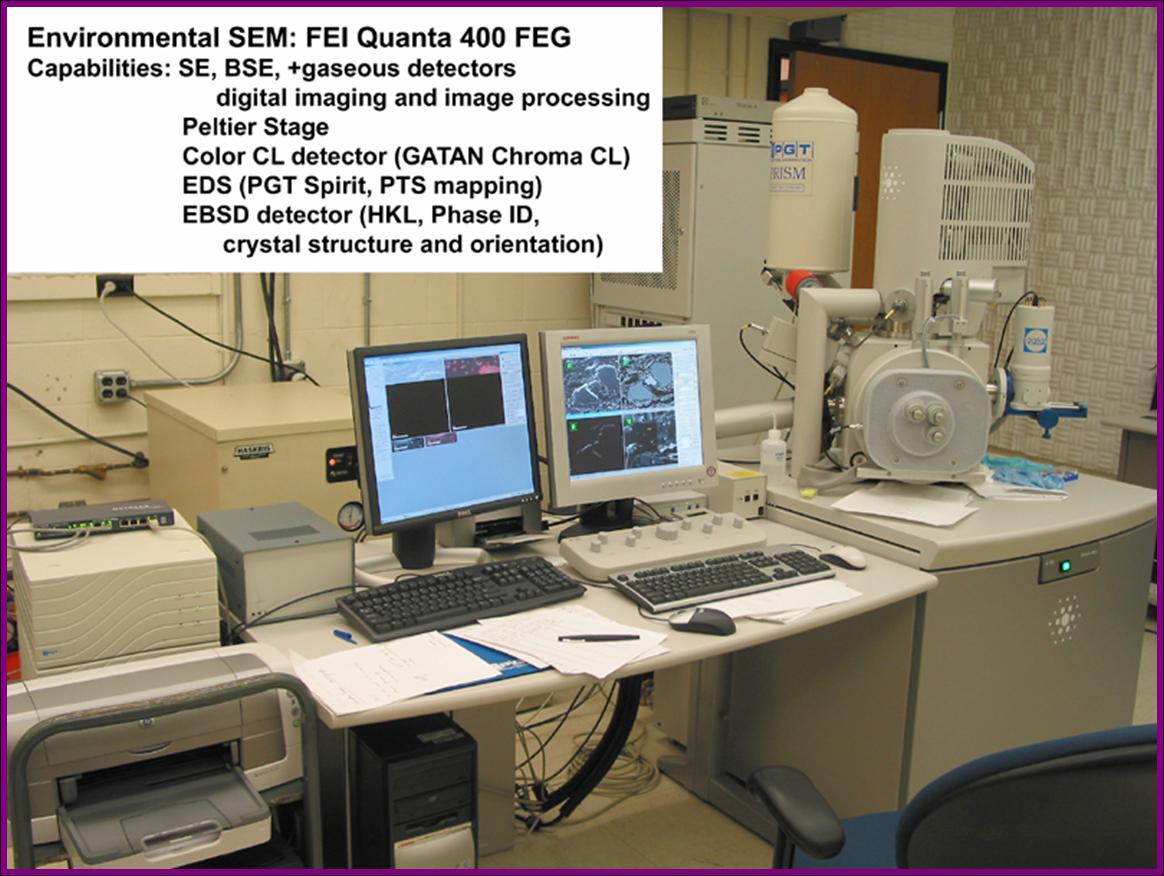
Our most powerful tool - "resistance is futile".
FEI Quanta 400 FEG (field emission environmental scanning electron microscope). Access to the nanometer realm, chemical characterization, crystal structure, grain orientation, high resolution color cathodoluminescence. No coating needed, can image moist materials, microbes survive study under the beam.
NSF funded.
Publications from SEM DATA
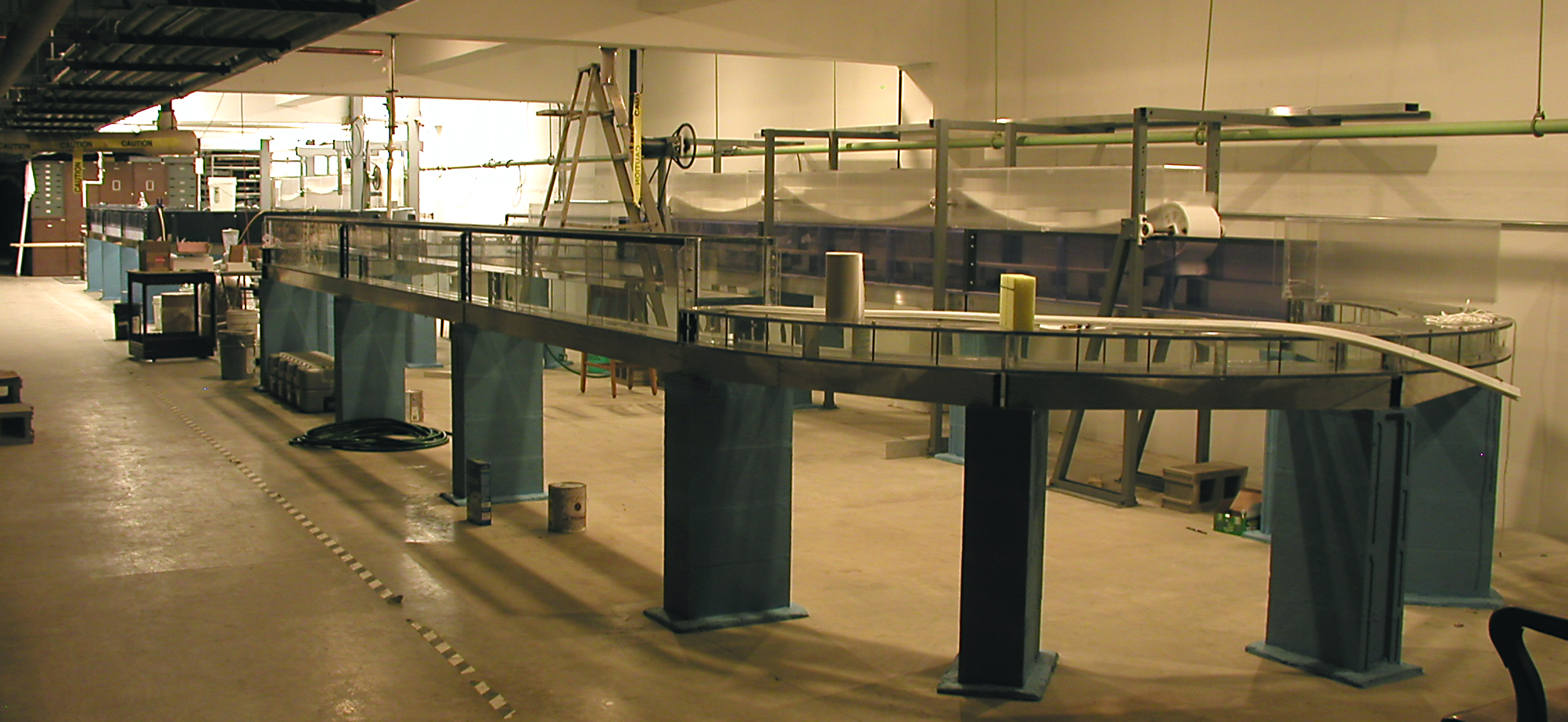
Where the fundamentals of mudstone sedimentology are explored.
This flume facility was built to study depositional and erosional parameters of mud, and to duplicate depositional features observed in the rock record (reverse engineering mother nature). It is a work in progress because we are constantly learning and building (4 flumes at present). NSF and industry funded.
View some results, SCIENCE Dec 14 2007, and our YouTube video.
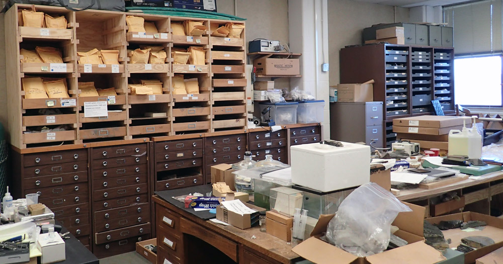
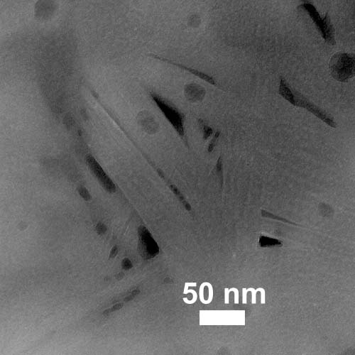
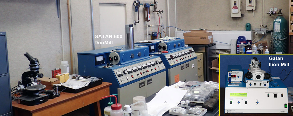
Where we make samples to give up their best held secrets.
Indispensible - Where it all starts.

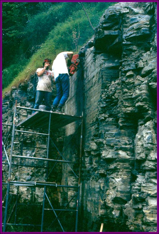
Unobscured textural relationships - most desirable samples.
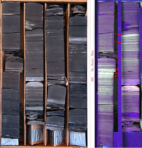
Next to detailed, cm by cm core descriptions, we also use observations with ultraviolet light to see hidden details of the shale record. We use a custom build UV bench to make UV observations and photography.
The pictured core (left) shows reworked horizons and bioturbated intervals. Adding UV photography (right, purplish image) shows concentrations of abundant algal cysts (marked with red arrows) that indicate sediment starved intervals.
We also own several teaching cores that have been preserved as board-mounted polished slabs.
Teaching shales and muds from Archean to Modern.
Thirty years of collecting shales do build up. Our collection of shale sample suites ranges from material as old as Archean and as young as a few years old (stabilized seafloor sediments). Our teaching collection consists of 20 pallets of samples and hand specimens, several thousands of polished slabs, about 3000 thin sections, hundreds of ion milled samples, and an extensive collection of petrographic microscope, SEM, and TEM images.