|
|
|
|
|
|
|
|
|
| Microbes are all around us. In
terms of numbers they rule the earth, and they manage to live under
extreme conditions, such as in hot springs, in polar ice, at depth
in rock formations, and in any conceivable environment in between.
Of interest for this paper are microbes that colonize, stabilize,
and modify the surfaces of sandy and muddy sediments. Microbial mat
deposits in carbonates are excluded, although the vast majority of
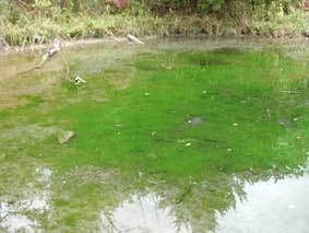
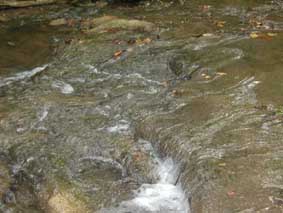
published examples is found in that rock type. Modern microbial mats on sandy sediment surfaces are well documented. The microbial colonization of muddy surfaces, although less known, can be readily observed in modern puddles a few days after a rainstorm. Essentially, for any sedimentary surface there is likely a microbial community that is equipped to thrive there if sedimentation rates are sufficiently small. Some can even prosper in absence of sunlight, or in the harsh and dry conditions of deserts. Considering how many times the "present has been the key to the past", it stands to reason that numerous ancient sand and mud surfaces were colonized by microbial communities. Thus, we may assume that sandstone and mudstone deposits of the geologic record should abound with microbial mat deposits It is at this point that an old geologic truth asserts itself once again: "just because something should have existed does not mean that it has been preserved". Positively proving the former presence of microbial mats in sedimentary rocks of any kind is actually quite a formidable task. Even in modern microbial mat systems, all remnants of the constructing microbiota may be destroyed within the first few hundred years of burial. Furthermore, if one applies strict criteria of biogenicity, such as preserved filaments in live position to published examples of fossil microbial mats (or stromatolites), only a very small portion can actually be shown to have a clearly demonstrable organosedimentary origin. So, what is a sedimentologist to do? Simply play it safe and assume that a given laminated sediment is not of microbial origin because proof positive is missing? Maybe we should look at the question from a different angle. In trace fossils, for example, the animal that produced the fossil is typically not preserved in association with the burrow. There is no dispute, however, that the traces were produced by a variety of animals, and that traces can be considered "fossilized behavior". As these animals were searching for food or seeking shelter, they invariably modified the sediment and produced trace fossils that record the interaction of the animal with the sediment. Likewise, laminated sediments produced by microbial mats can be considered the "trace" that reflects the interaction of the mat community with the environment. Microbial Mats in Mudstones (and sandstones) Unfortunately, laminated sediments can just as well be produced by purely physical sedimentation processes. Thus, there is a need to develop criteria for the identification of microbial mat laminae. The best place to start looking for microbial mat deposits in sandstones and mudstones is the Proterozoic. Its biosphere was dominated by microorganisms and the bulk of all published microbial mat deposits (stromatolites), albeit in carbonates, come from that time period. In the Phanerozoic, of course, bioturbation and grazing had a negative impact on the preserved biomat record. Nonetheless, if one starts looking around, one finds that even today, microbial films and mats abound in many sedimentary environments. In fact, it is hard to look at many aqueous environments without finding that sediment particles and sediment surfaces in many instances carry microbial communities. The resulting bio-films render the surface slippery (as most of us know from experience), bind particles together, and change the chemistry of the underlying sediments. Just for entertainment purposes I am showing in the space below microbial features that I noticed along a small stream near Sulphur, Oklahoma. Although streams are normally not thought of as suitable habitats for microbial mats, in this particular case we are dealing with a spring fed stream that has only a small suspended sediment load (would have negative impact on mat). Within this stream, and especially close to the origin of the springs, the entire stream bed may be covered with microbial covers that can reach several mm thickness, and may show morphologies reflective of flow conditions. |
|
|
|
|
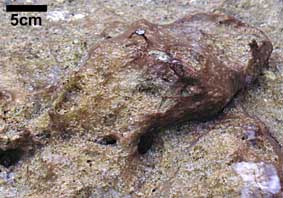
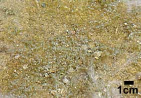
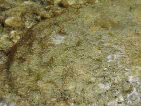
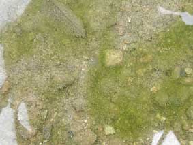
| Figure 1: Boulders and pebbles in creek bed are covered with a slippery film of microbial material. Gas bubbles (oxygen) are caught underneath the film. | Figure 2: Close-up of creek bed and microbial cover with bubbles. |
|
|
|
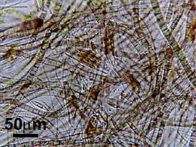
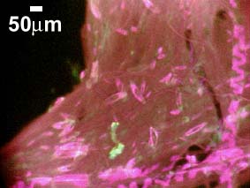
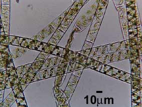
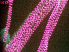
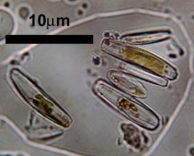
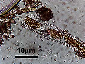
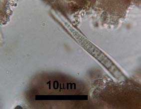
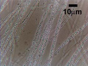
| Figure 3: The microbial cover is dominated by intertwined filaments of blue-green algae (Plankthotrix sp.?) and diatoms (Navicula sp. ?), as well as a moderate abundance of Spirogyra filaments. | Figure 4: A fluorescent light view of the microbial felt. Closely spaced blue-green algal filaments (low intensity fluorescence), Spirogyra at right and bottom (bright fluorescence), and diatoms (boat-shaped spots). |
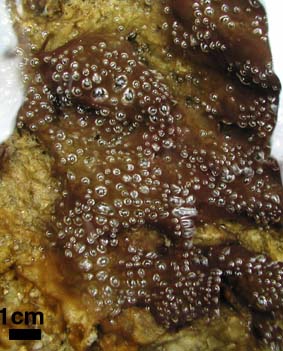
| Figures 1, 2, 5, and 6 show areas of rapid water flow, and microbial mats that more or less conform to the underlying substrate. In Figures 7 and 8 we see the effect of lower flow velocity. The microbial communities are able to develop vertical structures (phototactic response). | |
|
|
|
| Figure 5: Low relief mat covers boulders and sediment in swiftly flowing water. Darker spots are areas with higher diatom abundance. | Figure 6: Low relief mat in rapid flow area (note elongated bubbles). Dark color indicates abundant diatoms within the blue-green algal felt. |
|
|
|
| Figure 7: At somewhat slower flow vertical pinnacles form. These are buoyed by gas bubbles, but also firm enough to stand up without being buoyed. Darker colors indicate increased diatom abundance. | Figure 8: Abundant pinnacles of blue-green algae and diatoms in calmer water. Gas bubbles provide buoyancy. |
| The blue-green algal/diatom community illustrated above seems to have a general preference for running water. In stagnant water, such as the pool below, felts of green algae (Spirogyra) completely dominate the microbial bottom cover. | |
|
|
|
| Figure 9: Pool along Travertine Creek, bottom covered with felt of filamentous green algae. |
Figure 10: Close-up of green algal bottom cover. |
|
|
|
| Figure 11: Detail view of algal filaments. The large cell size indicates that it is a green algae, and the spiraling chloroplast indicates a species of Spirogyra. | Figure 12: Fluorescent light picture. The chloroplast fluoresces red because of photosynthetic pigments. |
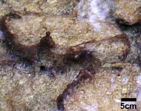
| Further down on along the creek, small rapids are characterized by smooth rounded bodies of calcium carbonate. | |
|
|
|
| Figure 13: Small rapids with rounded calcium carbonate deposits (tufa/travertine). | Figure 14: The slippery surface of the carbonate crust is dominated by diatoms. |
|
|
|
| Figure 15: Diatoms connected by a sheath of extracellular mucus. | Figure 16: Blue green algal filaments are also present. |
| In places where artesian springs have tapped reducing ground waters that carry hydrogen sulfide (rotten egg smell) and are flowing towards the creek, we may also find white slimy covers in the stream channels. | |
|
|
Figure 17: The white stuff consists of vegetation covered by white sulfur bacteria (soft slimy consistency) that gain their energy by oxidizing hydrogen sulfide. The pinkish hue could be due to the presence of sulfur purple bacteria. |
|
|
|
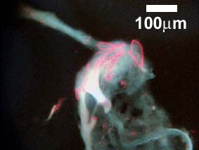
| Figure 18: Microscope photo of white sulfur bacteria. the granules within the filaments consist of sulfur. |
Figure 19: Clump of sulfur bacteria under blue light fluorescense. The red stringers could be the response of pigments in sulfur purple bacteria. |
| None of the above described microbial covers are likely to make it into the geologic record in a big way, but they nonetheless have a clearly noticable impact on presently operating sedimentation processes. This in short is the challenge for sedimentologists: how do we detect the effects of microbial mats and films that may have been a major part of ancient sedimentary environments? | |
|
|
|
| Back to SHALE RESEARCH LAB Main Page | |
| Back to IU Department of Geological Sciences | |
|
© Jürgen Schieber, IU Bloomington Department of
Geosciences Last updated: November 23, 2023. |
|